Volume Assessment… … is hard
It is hard for many reasons, one of which is terminology. Is the patient “dry“? Are they dehydrated (or more accurately hypertonic) or volume depleted (have decreased extracellular volume) [1]? What is the “volume status“?
One reason to make this assessment is to decided to give fluids, or volume expand. But then one needs to consider is the patient fluid responsive, or at the least fluid tolerant. And once that decision is made we will not even begin to consider the implications of the type of fluid given.
Traditional physical exam teachings for “volume” include looking at skin turgor or axillary sweat. Orthostatics are often used as a surrogate for volume. Capillary refill has been referred to as “useless” [2], though it has made a resurgence as a marker of end organ perfusion [3], not volume assessment. More recently static (less useful) and dynamic markers of volume responsiveness have been proposed, but these have variable utility depending on if the patient is spontaneously breathing or not and require some surrogate measurement of cardiac output for optimal performance [4, 5].
A brief look at some of the evidence for traditional physical exam:
Axillary sweat and Skin Turgor
Per Dr. McGee’s seminal Evidenced Based Physical Diagnosis the pooled operating characteristics for axillary sweat and skin turgor in detecting hypovolemia, defined here as hypertonicity, are as follows:
| Sensitivity (%) | Specificity (%) | +LR | -LR | |
| Dry axilla | 40-50 | 82-93 | 3.0 | 0.6 |
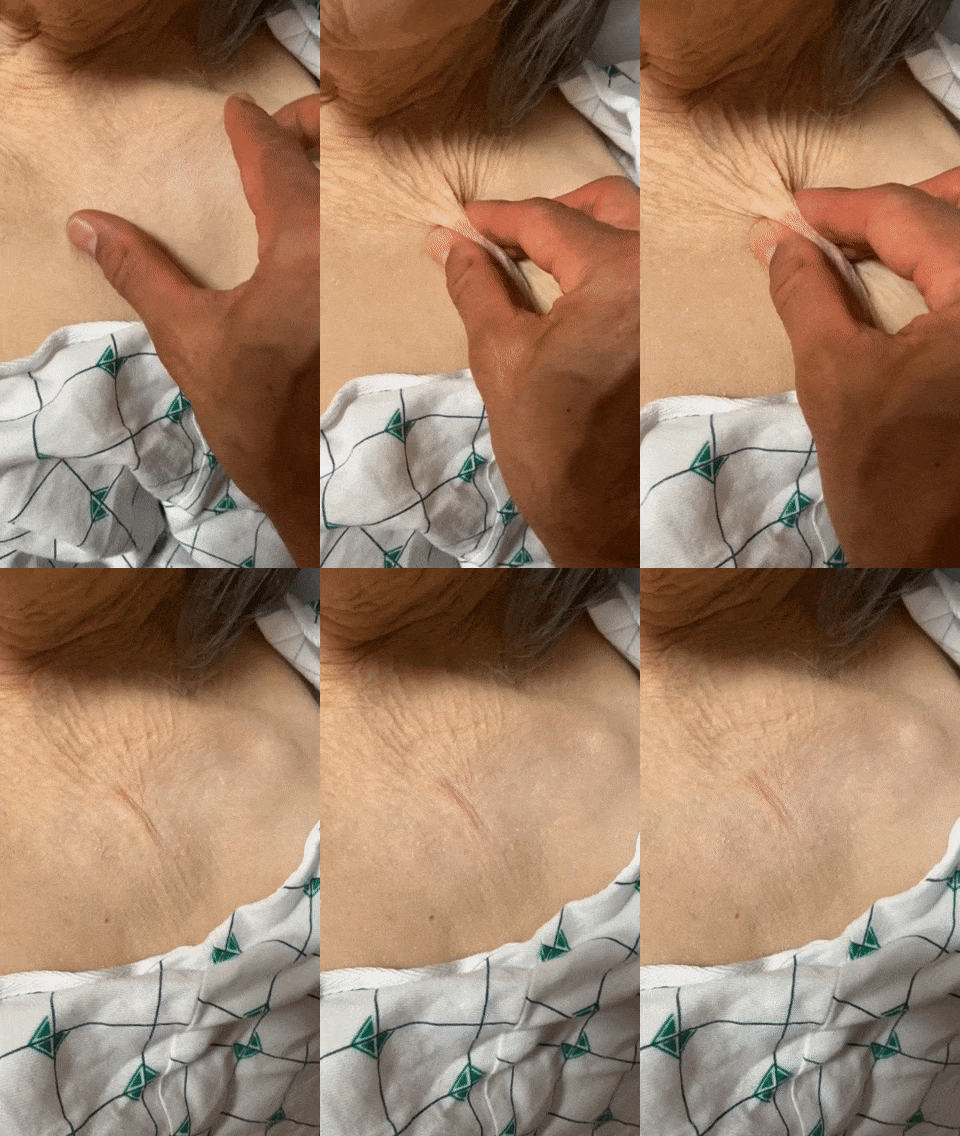
| Abnormal skin turgor (subclavicular area) | 73 | 79 | 3.5 | 0.3 |
It should be noted that other findings such as dry mucous membranes, tongue furrows and sunken eyes are often considered, but they are more subjective.
These are marginally good operating characteristics for hypertonicity. So how does one assess these and are they practical?
Axillary Sweat
Abnormal skin turgor
Orthostatics and Capillary Refill time
Orthostatics have not performed well for hypovolemia as defined above [9]. Early studies used healthy volunteers that were phlebotomized (i.e. blood loss, not hypertonicity) moderate (450-630 mL) to large (630 – 1150 mL) amounts of blood [6,10]. A ≥20 mm Hg decrease in SBP was only 9% sensitive for moderate blood loss! Better maybe actually a pulse increase ≥30/min or the subjective sensation of severe dizziness, but only for large blood loss.
| Moderate Blood Loss, Sensitivity (%) | Large Blood Loss, Sensitivity (%) | Specificity (%) | |
| Pulse ↑ ≥30/min or severe postural dizziness | 7-57 | 98 | 99 |
| ↓ SBP ≥20 mm Hg | 9 | — | 90-98 |
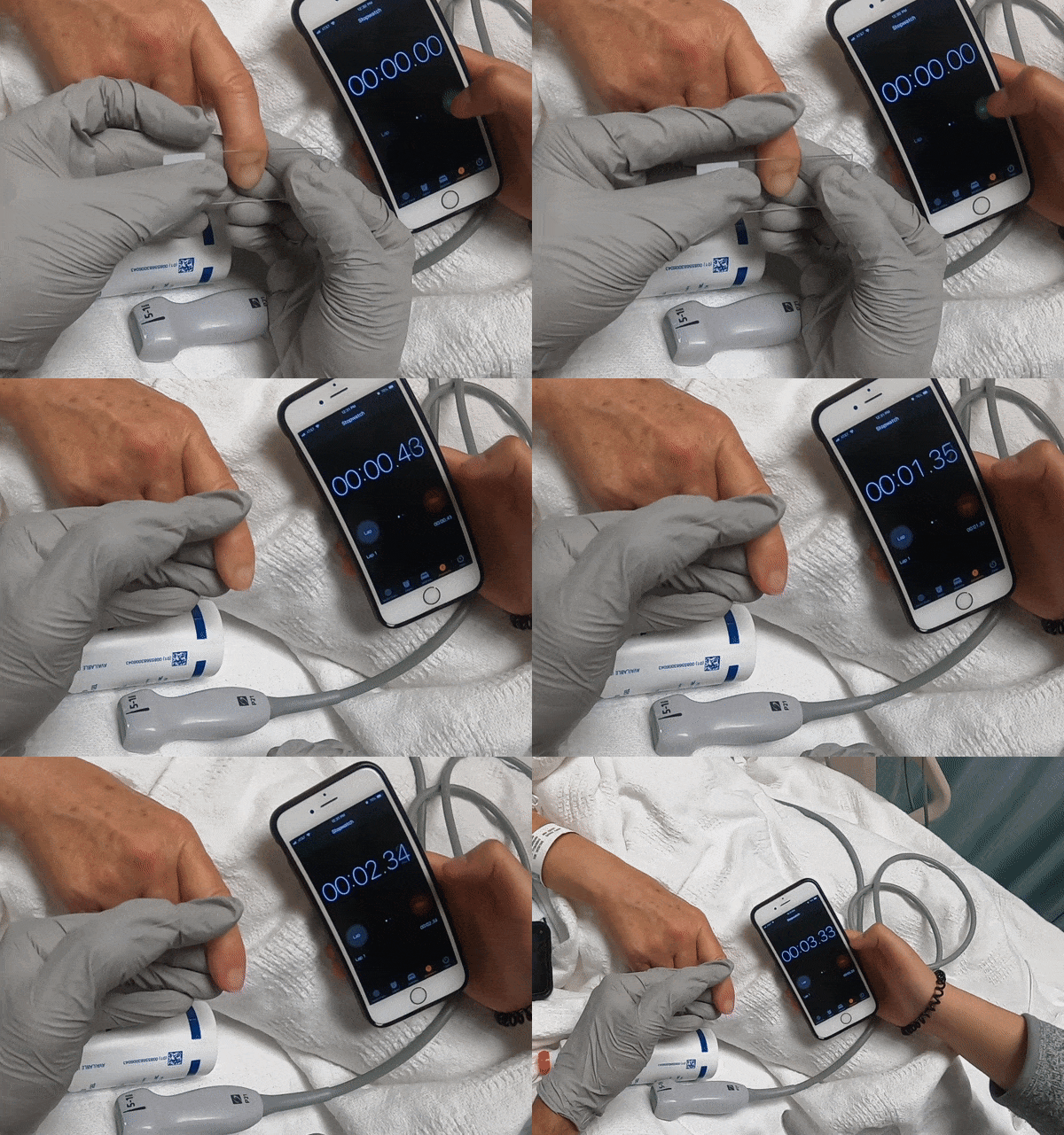
What about capillary refill (CRT), not for hypovolemia [2,10], but peripheral perfusion [3]? Is it practical and how do you standardize it?
CRT in ANDROMEDA-SHOCK was as follows:
- Apply firm pressure to the ventral surface of the right index finger with a glass microscope slide for 10 seconds.
- Normal skin color was registered with a chronometer, and a refill time >3 seconds was abnormal.
Volume Responsiveness
Turgor may be the only practical exam finding for hypovolemia that is helpful, though it is only modestly so. We are extrapolating when using orthostatics and capillary refill in hypovolemia, those are shown useful in other scenarios (large blood loss, tissue perfusion). Do turgor or the other findings discussed predict volume responsiveness? Sadly turgor does not, predict responsiveness, nor do the others [5]. The best option may be passive leg raise, but this depends on dynamic assessments of cardiac output (or surrogates) and have been discussed elsewhere (i.e. VTI 1, VTI 2).
- Bhave G, Neilson EG. Volume depletion versus dehydration: how understanding the difference can guide therapy. Am J Kidney Dis. 2011 Aug;58(2):302-9.
- Watson A, Kelly AM. Measuring capillary refill time is useless. Emergency Medicine Australasia. June 1993.
- Hernández G, Ospina-Tascón GA, Damiani LP, et al.; Effect of a Resuscitation Strategy Targeting Peripheral Perfusion Status vs Serum Lactate Levels on 28-Day Mortality Among Patients With Septic Shock: The ANDROMEDA-SHOCK Randomized Clinical Trial. JAMA. 2019 Feb 19;321(7):654-664.
- Monnet X, Marik PE, Teboul JL. Prediction of fluid responsiveness: an update. Ann Intensive Care. 2016 Dec;6(1):111. doi: 10.1186/s13613-016-0216-7.
- Bentzer P, Griesdale DE, Boyd J, MacLean K, Sirounis D, Ayas NT. Will This Hemodynamically Unstable Patient Respond to a Bolus of Intravenous Fluids? JAMA. 2016 Sep 27;316(12):1298-309.
- Stephen McGee’s Evidenced Based Physical Diagnosis, 4th ed, Elsevier, Philadelphia, PA, 2018.
- Eaton D, Bannister P, Mulley GP, Connolly MJ. Axillary sweating in clinical assessment of dehydration in ill elderly patients. BMJ. 1994 May 14;308(6939):1271.
- Kinoshita K, Hattori K, Ota Y, et al.; The measurement of axillary moisture for the assessment of dehydration among older patients: a pilot study. Exp Gerontol. 2013 Feb;48(2):255-8.
- Chassagne P, Druesne L, Capet C, Ménard JF, Bercoff E. Clinical presentation of hypernatremia in elderly patients: a case control study. J Am Geriatr Soc. 2006 Aug;54(8):1225-30.
- McGee S, Abernethy WB 3rd, Simel DL. The rational clinical examination. Is this patient hypovolemic? JAMA. 1999 Mar 17;281(11):1022-9.


